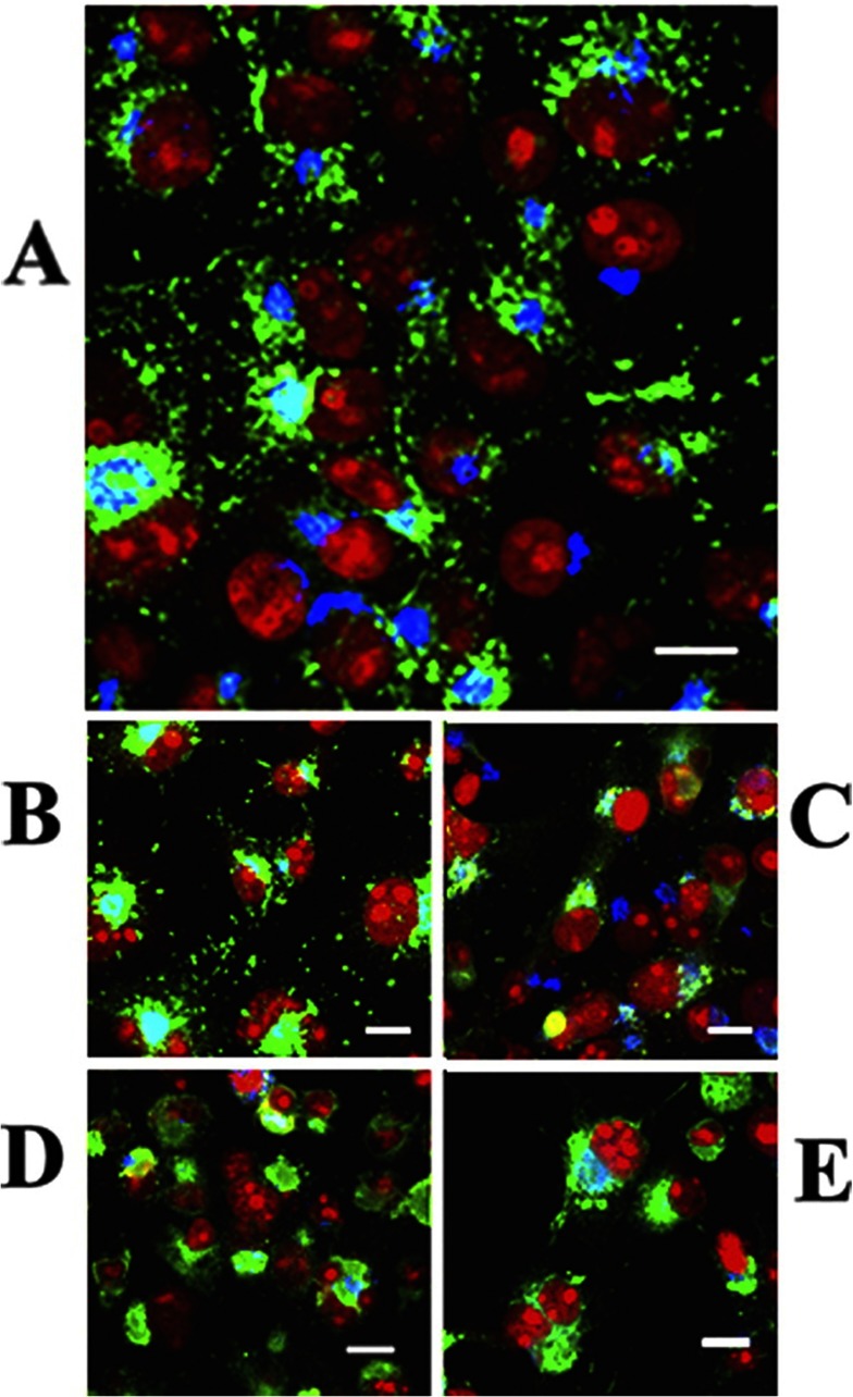
Figure 5. Intracellular distribution of ATP7B in COS-1 cells expressing WT ATP7B (A) or ATP7B subjected to mutations at Asp1027 (B), at Ser478, Ser481, Ser1121 and Ser1453 (C), at the transmembrane domain (TMBS) copper site (D) or at the sixth NMBD copper site (E).
Note the presence of cytosolic trafficking vesicles of WT ATP7B and even following Asp1027 mutation, but no trafficking following serine, NMBD or TMBS copper site mutations. All panels present different fields of cells treated identically. A copper load (200 μM) was added to the culture medium 2 h following infection with adenovirus vector for delivery of ATP7B cDNA (WT or mutant). The cells were fixed 24 h later. Secondary antibodies were goat anti-mouse conjugated with Alexa Fluor® 488 for the ATP7B c-Myc tag (green) and donkey anti-rabbit conjugated with Cy5 (indodicarbocyanine) for Golgi (blue). Red indicates nuclei stained with propidium iodide. Scale bar, 10 μm. This Figure was adapted from J. Biol. Chem. [60] Pilankatta, R., Lewis, D. and Inesi G. Involvement of protein kinase D in expression and trafficking of ATP7B (copper ATPase). Journal of Biological Chemistry. 2011; 286, 7389–7396.

