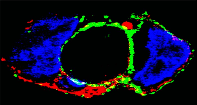
Figure 6. Association of ATP7A protein with the plasma membrane of COS-1 cells.
COS-1 cells were infected with optimal rAdATP7Amyc viral titres, and 200 μM copper was added. After 24 h, the cells were fixed with paraformaldehyde, permeabilized with Triton X-100, and stained. Immunostaining of ATP7A with the anti-Myc tag antibody is shown in green, and plasma membrane immunostaining with the anti-pan-cadherin antibody is shown in red. Nuclear staining with DAPI is shown in blue. Yellow indicates co-localization of ATP7A and anti-pan-cadherin antibodies in the plasma membrane. Scale bar, 10 μm. Reproduced from [61]. Copyright © 2010 American Chemical Society.

