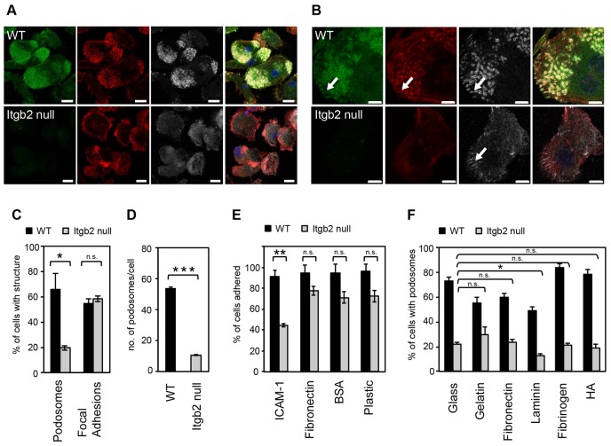
Fig. 1.
β2-integrin-null DCs are podosome deficient. Wild type (WT) and Itgb2-null SDCs plated on glass coverslips were fixed and stained for β2 integrin (green; FITC), F-actin (red; Alexa-Fluor-555) and vinculin (grey; Alexa-Fluor-633). (A) WT cells contained podosome clusters with clear actin cores, and β2 integrin- and vinculin-rich podosome rings and/or plaques. Itgb2-null DCs adhered but did not form podosomes. (B) WT and Itgb2-null DCs, can both form focal adhesions (white arrows). Single optical sections of 0.7 µm, taken at the ventral surface of the cells were acquired using Zen 2009 software on a Carl Zeiss 700 confocal laser-scanning microscope with a 100x Plan Apochromat/NA 1.46 oil immersion objective. Scale bars: 10 µm (A), 5 µm (B). (C) Percentage of integrin-null cells containing podosomes confirms the dramatic lack of podosomes compared to WT DCs (*P = 0.01, unpaired t-test), whereas the percentage of cells containing focal adhesions is normal. (D) Individual podosomes were also counted in WT and Itgb2-null SDCs and the results demonstrate that individual Itgb2-null cells have less podosomes compared with WT (29–57 cells scored per sample, error bars are s.e.m. for triplicate biological samples, ***P<0.001, unpaired t-test). (E) DCs were plated for 75 min on substrates as indicated and the % of adherent cells assessed after washing. Only when Itgb2-null cells were plated on β2 substrate, ICAM-1, was there a significant reduction in adhesion (**P = 0.002, unpaired t-test). (F) DCs were plated for 2 hours onto coverslips coated with gelatin, fibronectin, laminin or fibrinogen (all at 10 µg/ml), or with HA (100 µg/ml), and cells with podosomes quantified after staining for F-actin and vinculin. There was no significant rescue of podosomes in Itgb2-null DCs when plated on the various substrates compared with plating on glass alone, except when cells were incubated on laminin, in which case podosome levels were reduced rather than rescued (paired t-tests).

