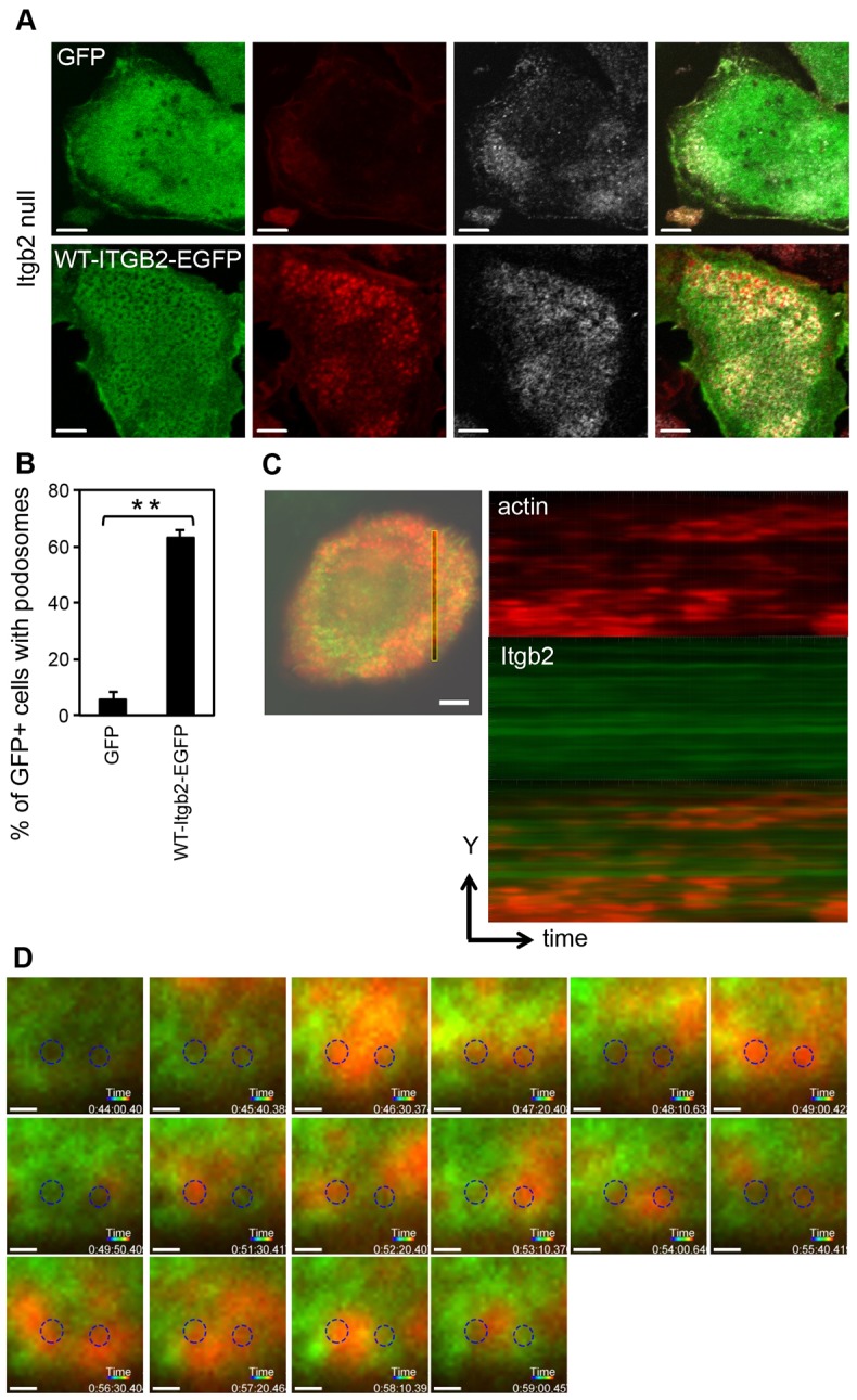
Fig. 3.

Retroviral expression of Itgb2-EGFP rescues podosomes in Itgb2-null DCs. Itgb2-null BMDCs were infected with retrovirus encoding a Itgb2-EGFP fusion protein, or GFP alone as a control. (A) The infected DCs were allowed to adhere to glass, then fixed and stained for F-actin (red; Alexa Fluor-555) and vinculin (grey; Alexa Fluor-633). Images were acquired using a Zeiss LSM700 as in Fig. 1. Scale bars: 5 µm. (B) Percentage of infected (GFP+) cells that contain podosomes, demonstrating significant reconstitution of podosomes with WT-β2 integrin compared to GFP alone (**P = 0.007, paired t-test). (C) Retrovirus encoding both Itgb2-EGFP and Lifeact-mCherry was used to infect Itgb2-null DCs. The cells were plated onto glass dishes and images collected every 10 seconds at 37°C using a Nikon Eclipse Ti TIRF microscope with an ApoTIRF 100x/NA1.49 objective as in Materials and Methods. A section through a podosome cluster was selected and Imaris software used to convert time to display on the z-axis, to generate a kymograph (Itgb2, green; actin, red; sequence represents 491 seconds), revealing the transient nature of the actin cores compared with the long-lived Itgb2-EGFP. To measure the lifetime of podosome cores, 682 podosomes were observed in six cells from at least three separate experiments. Scale bar: 5 µm. (D) A series of selected images (50-second intervals) shows that stable EGFP-labelled Itgb2 structures can support the reoccurrence of podosome cores in the same location after long periods of time (circled areas; images were cropped and circles were added to aligned layers using Photoshop CS5); ∼17±3% of sites (810 observed podosomes in six plaques from three experiments) hosted the return of actin cores. Scale bars: 0.5 µm.
