Fig. 4.
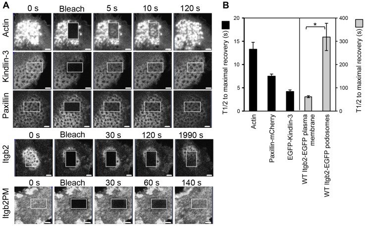
FRAP analysis of Itgb2-EGFP lifetime in podosomes. BMDCs were infected with retroviruses for expression of actin-EGFP, EGFP-kindlin3, paxillin-mCherry or Itgb2-EGFP (Itgb2-null cells). The cells were plated into glass-bottomed dishes and an area within a podosome cluster was photobleached using a Zeiss LSM700 confocal microscope as in described in Materials and Methods. Cells were then imaged over time to follow fluorescence recovery (A). For the Itgb2-EGFP-expressing cells an additional area of cell was also bleached to assess integrin turnover outside of podosomes (Itgb2PM). The fluorescence recovery in podosomes of actin-EGFP, EGFP-kindlin-3, paxillin-mCherry and Itgb2-EGFP in the plasma membrane were all relatively rapid compared to the recovery of Itgb2-EGFP in podosomes. (B) Recovery curves for each tagged protein were normalized for comparison and mean t1/2 values calculated, each from three independent experiments (three individual BMDCs cultures and viral infections), analyzing a minimum of ten cells per experiment (P = 0.04, paired t-test, comparing β2 integrin in podosomes versus plasma membrane). Scale bars: 2 µm.

