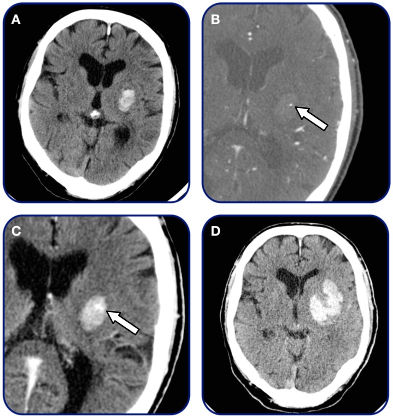
Figure 2.
Spot sign and contrast extravasation. Patient admitted within 2 h after symptom onset. (A) Initial non-contrast CT demonstrating small basal-ganglia hemorrhage, (B) CTA source image demonstrating single spot sign (arrow), (C) 3-min post-contrast CT-imaging demonstrating contrast extravasation (arrow), and (D) 24 h follow-up CT revealing final hematoma volume.

