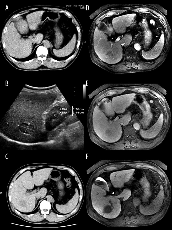
Figure 1.
Imaging of IHS in the patient. Abdominal plane computed tomography (CT) revealed a low-density mass in the posterior segment of the liver (C), whereas CT imaging 4 years previously detected no abnormal lesions (A). Ultrasonography revealed a hypoechoic heterogeneous mass 37×24 mm in size, with a hyperechoic rim in the right lobe of the liver (B). A dynamic study using gadoxetic acid-enhanced MRI revealed slightly inhomogeneous enhancement of the lesion in the arterial phase (D) and diminished enhancement in the equilibrium phase (E). In the hepatobiliary phase, the lesion become hypointense compared with the surrounding liver parenchyma (F).

