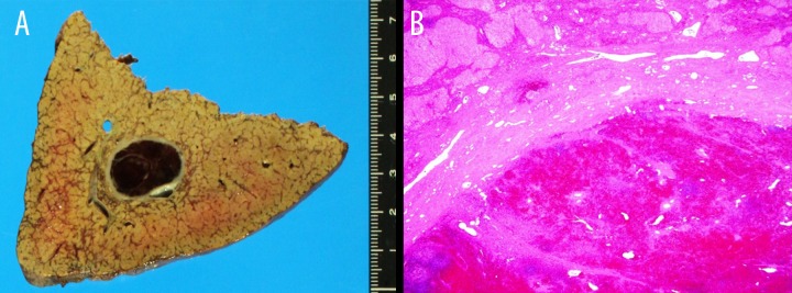
Figure 2.

(A) An encapsulated dark red mass was embedded in the cirrhotic liver parenchyma. (B) Hematoxylin and eosin staining of the lesion showed sinusoidal structures and lymphoid follicular aggregates with well-developed fibrous capsules with many small vascular channels.
