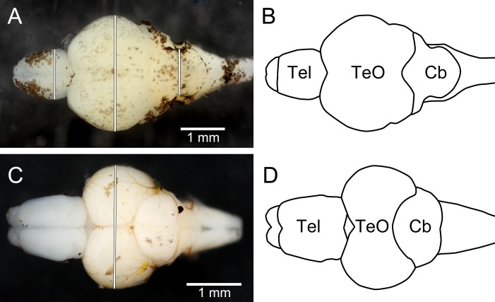
Figure 4. Dorsal photographs of brown trout fry brain (A), and juvenile zebrafish brain (C), with measurements taken marked as white lines. (B) and (D) show outlines of the trout brain and the zebrafish brain, respectively.
Tel, telencephalon; TeO, optic tectum; Cb, cerebellum. Rostral parts of the brains are pointing to the left.

