Figure 6.
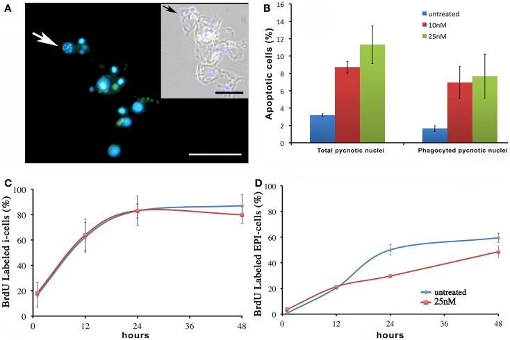
Effect of SiO2NPs on cell proliferation and death. (A,B) Cellular assessment of apoptosis induction by SiO2NPs. Following 24 h in 25 nM SiO2NPs, polyps were macerated and the percentage of apoptotic cells was determined by counting the DAPI-stained fragmented nuclei [arrow in (A)]. Scale bar: 50 μm. Inset in (A): phase contrast–DAPI merged of the same image. The arrow in the inset shows a pyknotic nucleus. The graph in (B) shows the percentage of total pyknotic nuclei and phagocytic moiety in normal and treated conditions. Cell cycling activity of interstitial stem cells (i-cells) (C) and of epithelial cells (EPI) (D) were estimated in untreated and 25 nM SiO2NPs treated animals by continuous incubation with BrdU, followed by maceration of 10 animals at the indicated time points followed by colorimetric immunostaining. SiO2NPs impaired the proliferating activity of epithelial, as shown by the lower percentage of BrdU labeled nuclei. In (B–D) data represent mean ± SD of three independent experiments; 100 cells were counted for each experiment.

