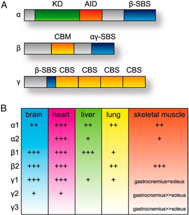
Figure 1.

Protein domains and tissue expression of AMPK subunits. A, The linear domain structure of the α, β, and γ subunits is shown. KD, kinase domain; AID, autoinhibitory domain; SBS, subunit binding sequence; CBM, carbohydrate binding motif; CBS, cystathione β-synthase motif. B, The relative protein expression of α, β, and γ isoforms in various organs is provided. Data are summarized from rodent models as previously described (11, 16, 157).
