Figure 2.

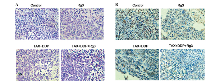
Histopathological and immunohistochemical assay of the tumor xenograft. (A) Hematoxylin and eosin staining. (B) The tumor cell proliferation was detected by Ki-67 staining (magnification, ×200). TAX, paclitaxel; DDP, cisplatin.

Histopathological and immunohistochemical assay of the tumor xenograft. (A) Hematoxylin and eosin staining. (B) The tumor cell proliferation was detected by Ki-67 staining (magnification, ×200). TAX, paclitaxel; DDP, cisplatin.