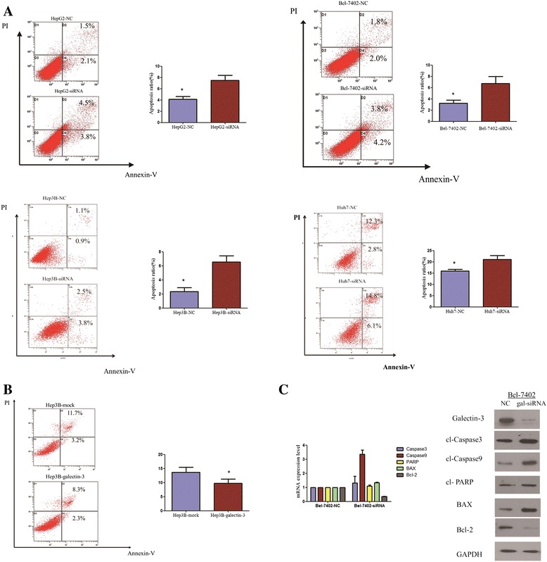
Figure 4.

Galectin-3 knockdown induced the apoptosis of HCC cells, and activates caspase-dependent apoptotic pathway. (A) Apoptosis levels of gal-siRNA and negative control-transfected HCC cells were quantified with an Annexin V and propidium iodide viability assay. After 72 hours, the cells were collected for apoptosis analysis. The percentages of Annexin-V or propidium iodide-positive cells were detected by flow cytometry. There were statistical differences between gal-siRNA cells and negative control cells. Experiments were performed in triplicate times (*P < 0.05, independent Student’s t-test). (B) The galectin-3 overexpression in Hep3B inhibited the apoptosis. Experiments were performed in triplicate times (*P < 0.05, independent Student’s t-test). (C) The protein and mRNA levels of PARP, caspase-3/9, BAX, and Bcl-2 proteins were analyzed by western blotting and RT-PCR.
