
Figure 5.
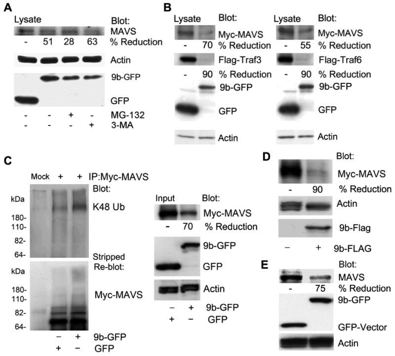
ORF-9b enhances MAVS proteasomal degradation via its K48-linked ubiquitination. (A) Immunoblots of lysates from HEK 293 cells expressing GFP-vector or 9b-GFP untreated or treated with MG-132 (10 μM) or 3-MA (5 mM) 4 h. The blots show the indicated proteins. (B) Immunoblots of lysates from HEK 293 cells expressing Flag-TRAF3 or Flag-TRAF6 and either GFP or 9b-GFP. The blots show the indicated proteins. (C) Immunoblots of Myc immunoprecipitates (+) and cell lysates from HEK 293 cells expressing Myc-MAVS and GFP or 9b-GFP. Following immunoblotting for K48 ubiquitin (Ub) the membrane was stripped and probed for Myc-MAVS. Mock is a control antibody immunoprecipation. (D) Immunoblots of lysates from HEK 293 cells expressing Myc-MAVS in the presence of either the Flag-vector or 9b-Flag. The blots show the indicated proteins. (E) Immunoblots of lysates from THP-1 cells permanently expressing GFP or 9b-GFP for endogenous MAVS. Also shown are GFP, 9b-GFP, and actin levels.