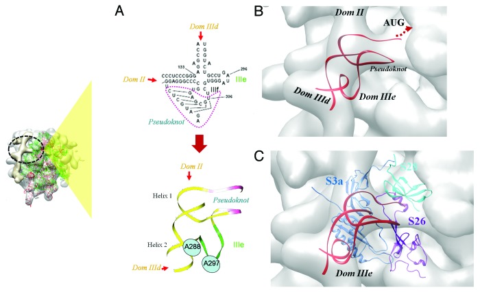
Figure 5. IRES-40S contact at the ribosomal platform region. (A) The secondary structure and 3D model of the HCV IRES comprising domains IIIef and pseudoknot. The position of two bases A288 and U297 that are reported to base-pair18,75 are highlighted. (B) Model of the IIIef+pseudoknot generated by interactive manipulation and structure refinement. The model fitted in the 40S platform density (gray) is shown. Locations of IIId, IIIe and II are also indicated (C) The docked model of IIIef+pseudoknot (red) and 40S ribosomal proteins S26 (purple), S3a (S1e) (blue), fitted in the cryoEM density (gray). The relative location of S28 (cyan) is also shown. The site of interaction involving domain IIIef+pseudoknot with respect to the complete ribosome, is indicated by the inset on the left.

An official website of the United States government
Here's how you know
Official websites use .gov
A
.gov website belongs to an official
government organization in the United States.
Secure .gov websites use HTTPS
A lock (
) or https:// means you've safely
connected to the .gov website. Share sensitive
information only on official, secure websites.
