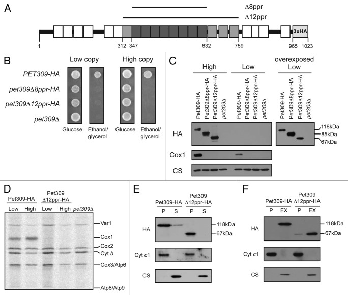
Figure 3. Pet309∆12ppr-HA is a peripheral inner membrane protein and is impaired for COX1 mRNA translation. (A) Diagram representing Pet309 as in Figure 2. The 12 PPR central motifs that were deleted are indicated in gray boxes. The 8 PPR motifs that were deleted in Figure 2 are shown in dark gray boxes. Residue numbers are as indicated. (B) Δpet309 cells were transformed with low-copy or high-copy number plasmids containing the wild-type PET309-HA, the pet309Δ8ppr-HA, the pet309Δ12ppr-HA genes or empty vector (pet309Δ). Cells were spotted on synthetic complete medium lacking uracil with glucose or ethanol/glycerol, and incubated for 4 days at 30 °C. (C) 10 μg of mitochondrial protein from the wild-type (PET309-HA), pet309∆8ppr-HA, and pet309∆12ppr-HA constructs expressed from high-copy or low-copy number plasmids were analyzed by SDS-PAGE and western blot. The membrane was probed with anti-HA, anti-Cox1 or anti-citrate synthase (CS) antibodies. In order to visualize the HA signal from the low copy plasmids, the blot was overexposed (right panel). (D) Wild-type, pet309∆12ppr-HA and pet309∆ cells were labeled with 35S methionine in the presence of cycloheximide for 15 min at 30 °C, the proteins were analyzed by SDS-PAGE and autoradiography. Cytochrome c oxidase subunit 1, Cox1; subunit 2, Cox2; subunit 3, Cox3; cytochrome b, Cytb; ATPase subunit 6, Atp6; subunit 8, ATP8; subunit 9, Atp9; ribosomal protein, Var1. (E) 100 μg of mitochondrial protein from the wild-type and pet309∆12ppr-HA strains were sonicated and centrifuged to separate the membrane (P) and soluble (S) fractions, and were analyzed by western blot using antibodies to HA, citrate synthase as a soluble protein marker, and cytochrome c1 as a membrane protein control. (F) 100 μg of mitochondrial protein from the strains in (E) were incubated with alkaline, 100 mM Na2CO3. The integral membrane (P) and soluble, extracted (EX) proteins were analyzed by western blot as in (E).

An official website of the United States government
Here's how you know
Official websites use .gov
A
.gov website belongs to an official
government organization in the United States.
Secure .gov websites use HTTPS
A lock (
) or https:// means you've safely
connected to the .gov website. Share sensitive
information only on official, secure websites.
