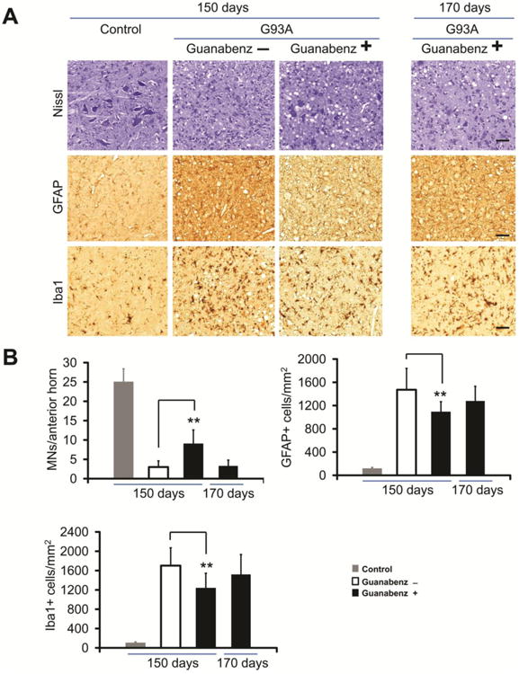
Figure 2.

Immunohistochemical studies of the anterior horn of the lumbar spinal cord of guanabenz-treated G93A transgenic mice, untreated G93A transgenic mice, and non-transgenic littermate mice at varying ages. Representative sections of the anterior horn of the lumbar spinal cord were stained with: (A) Nissl for MNs, anti-GFAP antibody for astrocytes, and anti-Iba1 antibody for microglia. (B) Bar diagrams show mean ± standard deviation calculated by counting cells in 15 to 20 sections from the lumbar spinal cord anterior horn of 4 mice from each of the following groups (guanabenz-treated G93A mice, untreated G93A mice, and littermate non-transgenic mice) at each age. The scale bar = 100μm. ** P < 0.001. The data were statistically analyzed using a t-test.
