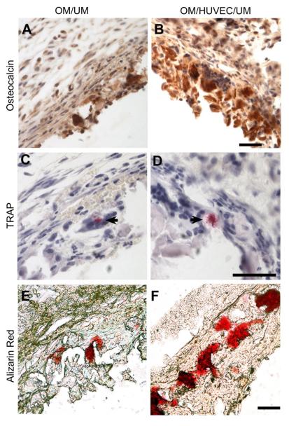
Fig. 9.
Immunohistochemistry staining images of osteocalcin for OM/UM (A) and OM/HUVEC/UM constructs (B). Osteoclast activity is shown through TRAP staining for in OM/UM (C) and OM/HUVEC/UM constructs (D) (black arrow). Alizarin red staining for mineralized calcium nodes in OM/UM (E) and OM/HUVEC/UM constructs (F) (Scale bar= 50 μm).

