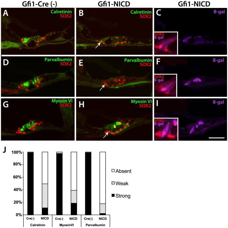
Figure 2. At P20, NICD-expressing hair cells in the cochlea have downregulated hair cell markers.
A–I Paraffin sections through the P20 cochlea stained for hair cell markers, SOX2, and ß-galactosidase. A, D, G. Calretinin, parvalbumin, and myosin VI all show expression in the inner and outer hair cells at P20 (although calretinin expression is weak in outer hair cells at P20). B, E, H. Both calretinin and parvalbumin are shut off in the Gfi1-NICD inner and outer hair cells, whereas myosin VI is generally still expressed in the outer hair cells in the mutant (H). Gfi1-NICD inner and outer hair cells express SOX2 and the inner hair cells have a more basally-located nucleus than in controls (B,E,H, arrows and insets in C,F,I showing higher power images of inner hair cells expressing ß-galactosidase and SOX2). J. Quantification of relative expression levels of indicated markers in the Gfi1-NICD (NICD) inner hair cells and littermate controls [Cre(-)]. Scale bar in I = 100 microns ( = 50 microns for inset panels).

