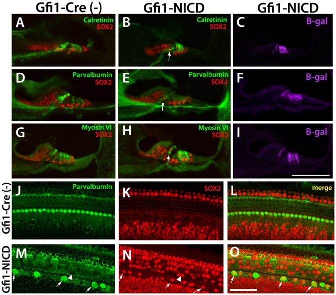
Figure 3. At P6, Some hair cell markers are downregulated in the inner hair cells.
A–I. Frozen sections through the P6 cochlea stained for hair cell markers, SOX2, and ß-galactosidase. A, D, G. Calretinin, parvalbumin, and myosin VI all show expression in the inner and outer hair cells at P6 (although parvalbumin expression is weaker in outer hair cells at P6). B, E, H. Both calretinin and parvalbumin are downregulated in the Gfi-NICD inner hair cells, although outer hair cell expression is largely maintained. Interestingly, myosin VI (G–I) is generally indistinguishable from the controls at this time point. Scale bar in I = 100microns. J–O. Wholemount cochlea stained for hair cell (parvalbumin) and supporting cell (SOX2) markers. Many of the inner hair cells have shut off parvalbumin and upregulated SOX2 by this time point (M–O, arrows). A few outer hair cells have downregulated parvalbumin (M–O, arrowhead), but most have upregulated SOX2.

