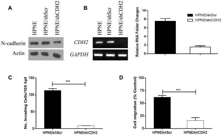
Figure 5. Invasion and migration assay after CDH2 knockdown in HPNE, HPNE/shScr, and HPNE/shCDH2 cells.
(a) N-cadherin was suppressed though shCDH2. Actin was used as the loading control. (b) RT-PCR (left) and real-time PCR (right) confirmed that CDH2 mRNA was significantly decreased in HPNE shCDH2 cells. (c) Modified Boyden chamber assay was performed with transfected cells. Cells were treated with or without 10 ng/ml TGF-β in serum-free medium in the top inserts, and 20% FBS medium was used in the bottom chamber as a chemoattractant. Invasive cells were counted in 3 fields at 10× magnification in duplicated inserts. (d) Wound-healing assay was performed with transfected cells treated with or without 10 ng/ml TGF-β. Three random images (4× magnification) were taken at the time of the scratch (0 hours) and at 20 hours. Migration rate was determined as the ratio of the distance traveled at 20 hours versus 0 hours in the wound's gap using Adobe Photoshop software. **P<0.01, ***P<0.001.

