
Figure 2.
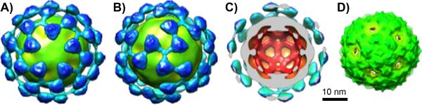
Cryo-electron microscopy image reconstructions at 17.4 Å resolution. (A,B) Qβ(mCherry)(Tfn)55 conjugate; views down the 5- and 3-fold symmetry axes, respectively. (C) Cross-sectional view showing added density other than that of the VLP in light blue. (D) Wild-type Qβ and Qb(mCherry).