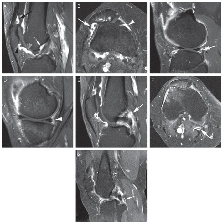
Figure 1.
Eleven anatomical sites evaluated in the proposed scoring system. (A) Sagittal T1-weighted contrast-enhanced (CE) image at the location of the anterior cruciate ligament (ACL). Definition of the suprapatellar site: 0.5–1 cm cranial to the superior patellar pole (black arrow). Definition of infrapatellar site: directly adjacent to the inferior patellar pole (black arrowhead). Definition of the intercondylar site: at the surface of Hoffa’s fat pad 1.5–2 cm distal to inferior patellar pole (grey arrowhead). Definition of ‘adjacent to the ACL’ site: directly anterior to the ACL close to its femoral attachment (white arrow). (B) Axial T1-weighted CE image at the location of the maximum medial-lateral patellar diameter. Definition of the medial parapatellar site: 0.5–1 cm posterior to the medial patellar pole. Definition of the lateral parapatellar site: 0.5–1 cm posterior to the lateral patellar pole. (C) Sagittal T1-weighted CE image at the location of the tibiofibular joint. Definition of the lateral parameniscal site: directly adjacent posterior to the posterior horn of the lateral meniscus (white arrow). (D) Sagittal T1-weighted CE image at the location of the tibial semimembranosus attachment. Definition of the medial parameniscal site: directly adjacent to the posterior horn of the medial meniscus (white arrowhead). (E) Sagittal T1-weighted CE image at the location of the femoral posterior cruciate ligament (PCL) attachment. Definition of ‘adjacent to the PCL’ site: Directly adjacent to the PCL at its mid-portion (white arrow). (F) Axial T1-weighted CE image at the location of the Baker cyst with peripheral enhancement indicating synovitis (white arrow). (G) Sagittal T1-weighted CE image showing a loose body (white arrow), located posteriorly to the PCL, surrounded by enhancing synovitis.

