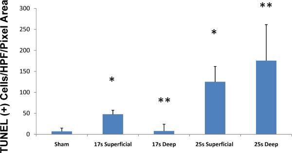
Figure 2. The full-thickness scald burn model markedly increases dermal tissue cell death.
Skin from sham, 17s and 25s burn animals was sectioned for fluorescein-labeled TUNEL as described. Sections were randomized; per section, 3 hair follicles were randomly selected, the ROI was defined and analyzed according to the described protocol using digital image software. A significant increase in cell death was seen in the 25 s burn group vs. the 17s burn group at both superficial (dermal/subcutis junction) and deep (subcutis/subQ muscle fascia) levels. Values are expressed as mean ± 95% confidence interval (* comparing the 25 s to 17 s superficial and ** comparing the 25 s to 17 s deep levels, p<0.001 for both comparison, ANOVA, n=15 HPF per group).

