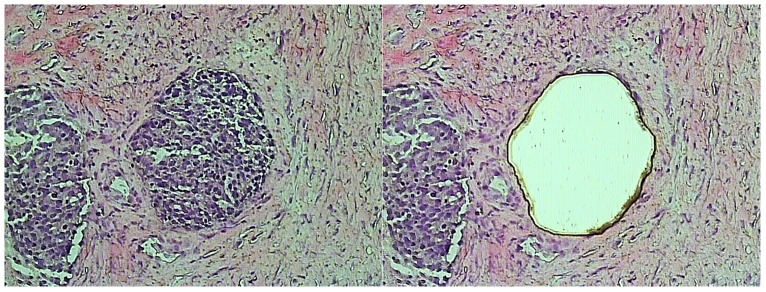
Figure 1. Laser capture microdissection of tumor tissue.

A 10 µm hematoxylin stained cryostat section of a serous adenocarcinoma of the ovaries. The picture on the left is prior to microdissection, on right the same slide after microdissection is shown.
