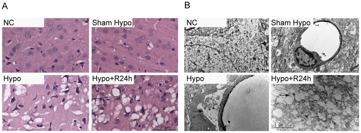
Figure 2. Edematous changes in hypoglycemic rat parietal cortex.
A. Hematoxylin and eosin staining showed that vacuolization around neuronal cell bodies and processes was evident after iso- EEG 60 min induced by hypoglycemia (Hypo) and recovery for 24 h(Hypo+R24 h). Nuclear are triangular. B. The end-feet around the capillaries were swollen (arrows), as were the mitochondrion (arrowheads) (Hypo). After 24 h recovery, edema was much more evident around the processes (Hypo+R24 h).

