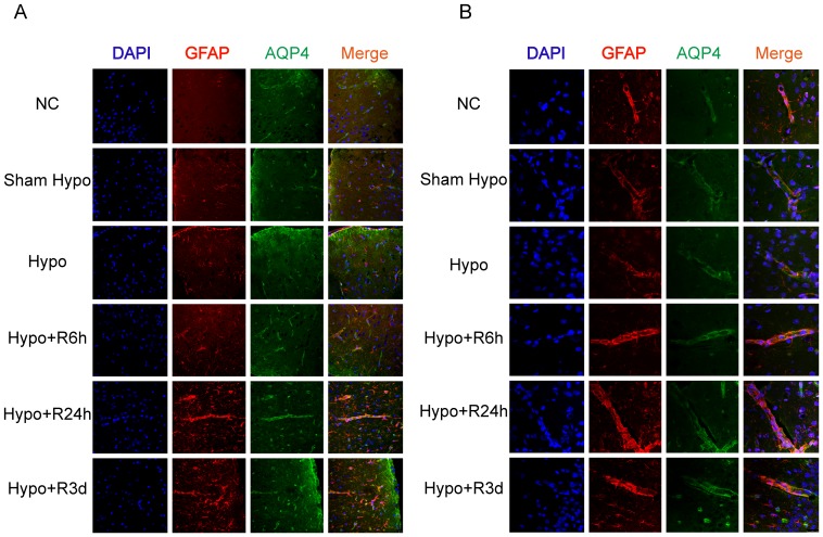
Figure 5. Increased immunereactivity of AQP4 in hypoglycemic rat brain.
A. Immunofluorescence staining for AQP4 showed strongest intensity in the rat cortex after 24-hour recovery from profound hypoglycemia. Reactive astrocytes were demonstrated more processes and stronger intensity of GFAP after hypoglycemia shock. B. Immunofluorescence staining for AQP4 showed stronger intensity on the end-feet around cerebral vessels in hypoglycemia group and recovery groups compared with normal control and sham hypoglycemia group.

