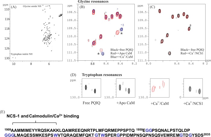
Figure 3.
Delineation of binding of NCS-1 to PQ 1909–2035. 1H–15N HSQC spectra of 15N-labeled PQ1909–2035 in the presence (A) and absence of CaM (B) and of NCS-1 (C), showing the glycine amide resonance, and (D) tryptophan NεH resonances. Panel A shows the full 15N–1H spectrum; expanded regions show resonances from only glycine amide (B and C) and tryptophan indole (D) groups. (E) The amino acid sequence of PQ 1909–2035 is shown with glycine and tryptophan residues colored blue. The likely CaM- and NCS-1-binding region is indicated.15

