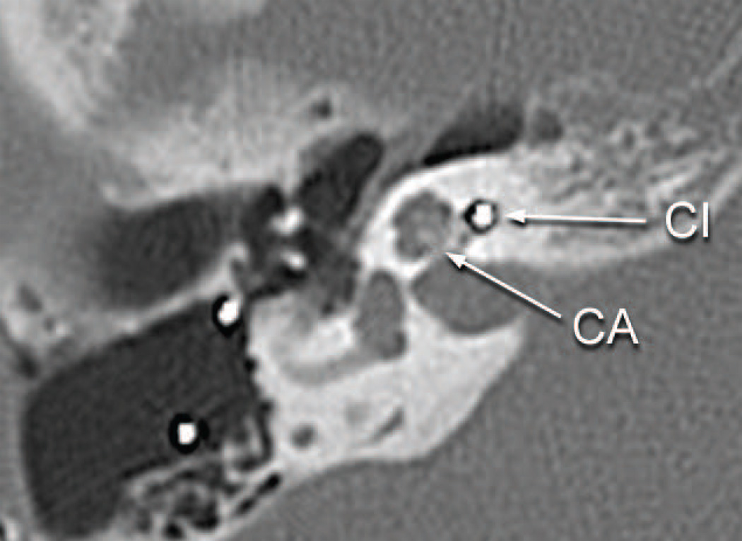
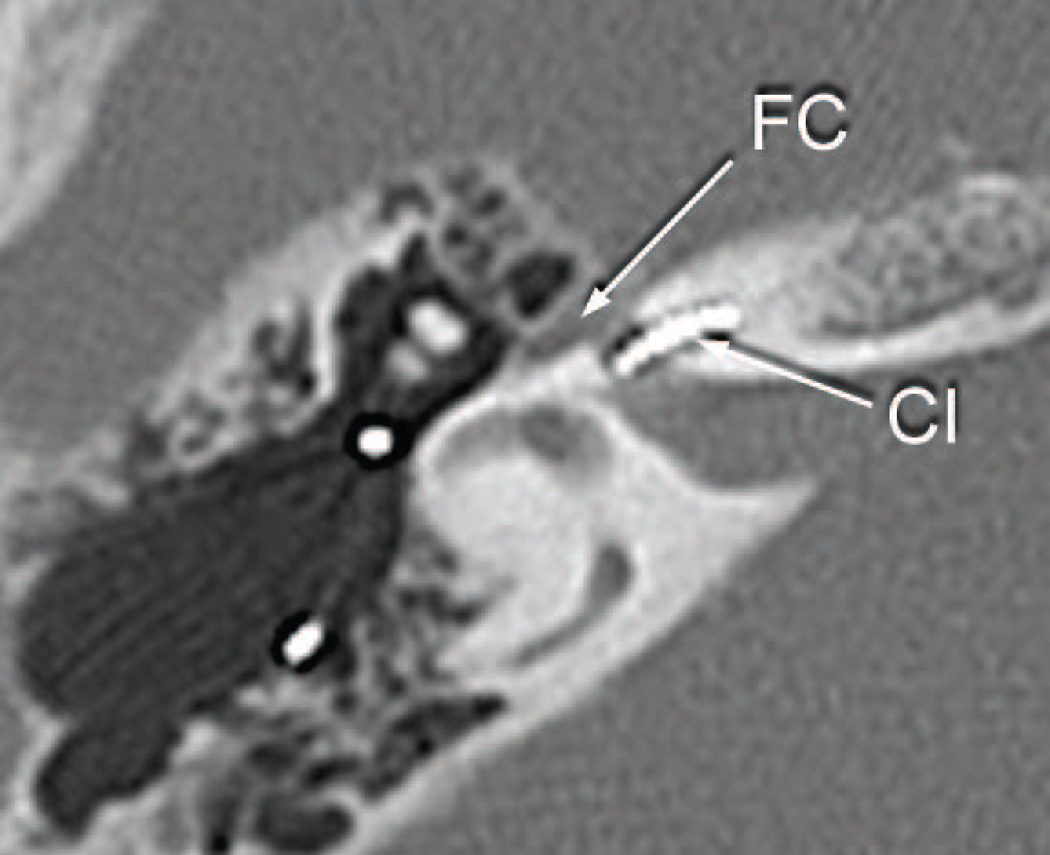
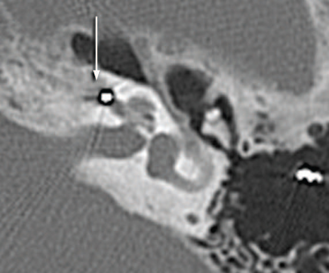
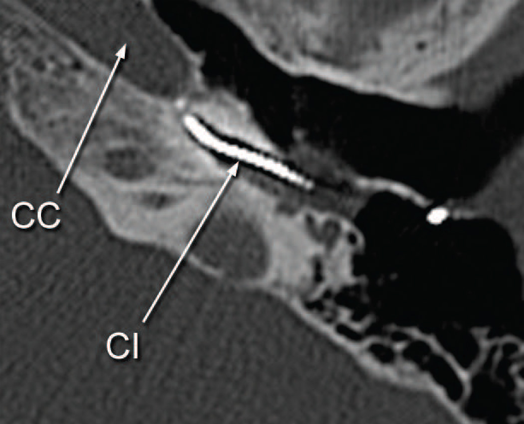
Fig. 3.
Postoperative CT scan of patient presented as a case report.
a. Right ear eight years following cochlear implantation and five years before death. The electrode array (CI) was seen in the basal turn. No abnormality of the cochlea was identified. The bone of the cribrose area (CA) was intact.
b. Right ear eight years following cochlear implantation. The facial canal in its horizontal segment (FC) was of normal caliber near the electrode of the cochlear implant (CI) in the basal turn.
c. Left ear six years following cochlear implantation, explantation and reimplantation and five years before death. An area of demineralization of the cochlear capsule medial to the basal turn was seen (arrow). The basal diameter of the basal turn of the cochlea was expanded anteriorly.
d. Left ear six years following cochlear implantation. The electrode (CI) was within the basal turn of the cochlea near the carotid canal (CC).


