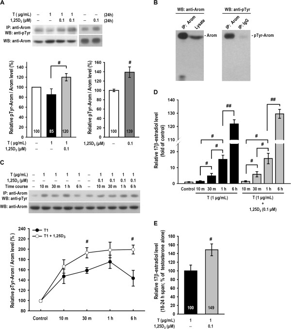
Figure 2.

1,25D 3 enhanced testosterone-induced aromatase tyrosine phosphorylation and 17β-estradiol secretion during 18–24 h. (A) Rat ovarian granulosa cells were treated with vehicle, testosterone, testosterone plus 1,25D3 or 1,25D3 alone for 24 h (n = 4–5 in each group). Aromatase was immunoprecipitated before being subjected to SDS-PAGE for resolution. The resultant blot was probed using both phosphotyrosine and aromatase antibodies. The expression of aromatase tyrosine phosphorylation (denoted by pTyr-Arom) was normalized to the corresponding aromatase expression and is represented as a percentage relative to the vehicle-treated control. (B) The left panel is a positive control that was both immunoprecipitated and immunoblotted with an anti-aromatase antibody. The lysate was also immunoprecipitated with the anti-aromatase antibody or IgG and immunoblotted with an anti-phosphotyrosine antibody, as shown in the right panel. A weak band in the IgG lane is as a negative control. (C) Cells were treated with testosterone alone or testosterone plus 1,25D3 as indicated for various time periods (n = 4–5 in each group). Each pTyr-Arom level was normalized using the corresponding aromatase level and is represented as a percentage relative to the vehicle-treated control. (D) After treatment for various time periods as indicated, the 17β-estradiol production levels were measured by radioimmunoassay. (E) After treatment with testosterone alone or testosterone plus 1,25D3 for 18 h, the culture medium was removed and then replaced with fresh medium. Cells were continuously treated for another 6 h (18–24 h span). After the final 6 h, medium was collected and the 17β-estradiol secretion levels were measured by radioimmunoassay (n = 6 in each group). The values represent the means ± SEM. #, p < 0.05; ##, p < 0.005 for comparison of the two treatment groups. The values listed within column indicate the means.
