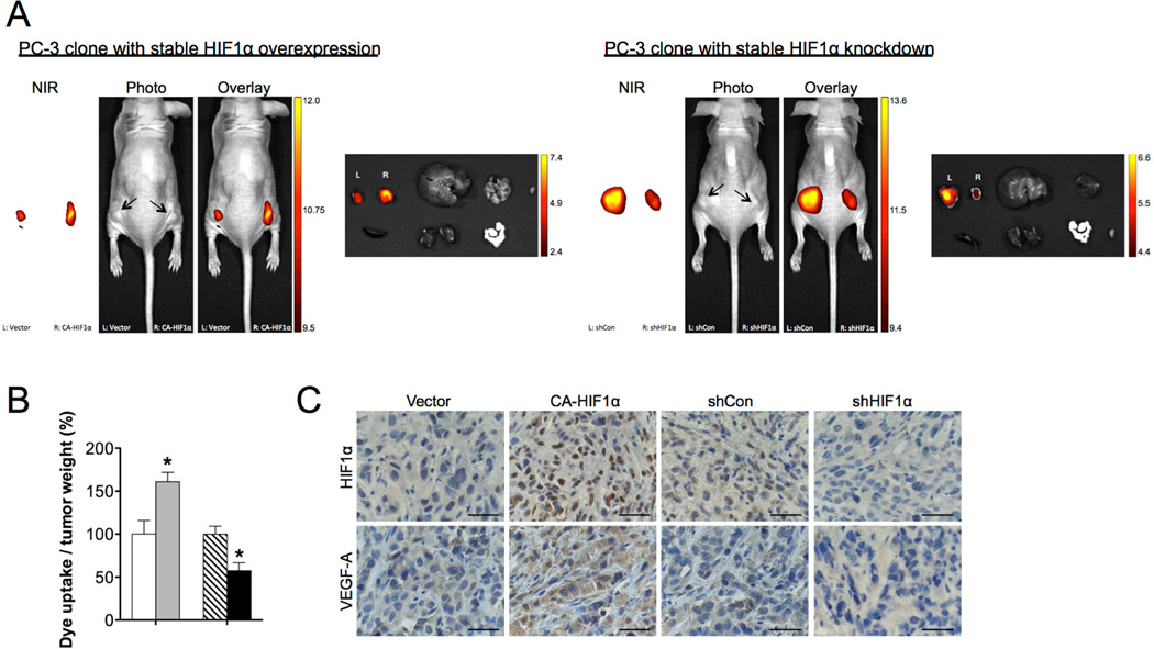
Fig. 3.
Hypoxia and HIF1α mediated the uptake of MHI-148 dye by tumor xenografts. (A) Representative in vivo and ex vivo NIRF images of control (left flank) vs. HIF1α-overexpressing (right flank) (left panel) and control (left flank) vs. HIFIα-knockdown (right flank) (right panel) PC-3 tumor xenografts. Scale bars represent x108 for both in vivo and ex vivo NIRF in the unit of radiant efficiency. (B) Quantitation of tumor uptake of MHI-148 dye (N=5, mean ± SEM) presented as the ratio of dye intensity to tumor weight. *p<0.05, **p<0.01. (C) IHC analysis of HIF1α and VEGF-A expression in PC-3 tumor xenografts with different manipulation of HIF1α levels as indicated. Representative images are shown. Original magnification, x400; scale bars represent 20 µm.

