
Figure 2.
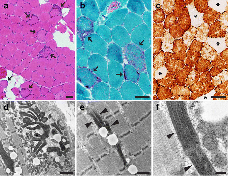
Paraspinal muscle pathology. Histochemical and ultrastructural analysis of T8 paraspinal muscle demonstrated evidence of a mitochondrial myopathy. (a) H&E stain (arrows indicate fibers with increase in basophilic staining indicative of mitochondrial hyperplasia), (b) modified trichrome stain (arrows indicate ragged red fibers), (c) COX (cytochrome c oxidase) stain (asterisks mark COX-negative fibers). Electron microscopy (d-f) shows mitochondrial hyperplasia, variation in the mitochondrial shape and size, and mitochondria with crystalline arrays (arrowheads). Of note, the crystalline arrays present in this case do not have a common “parking lot” appearance. Scale bars: a-c, 50 µm; d, 1.5 µm; e, 1 µm; f, 200 nm.