
Figure 1.
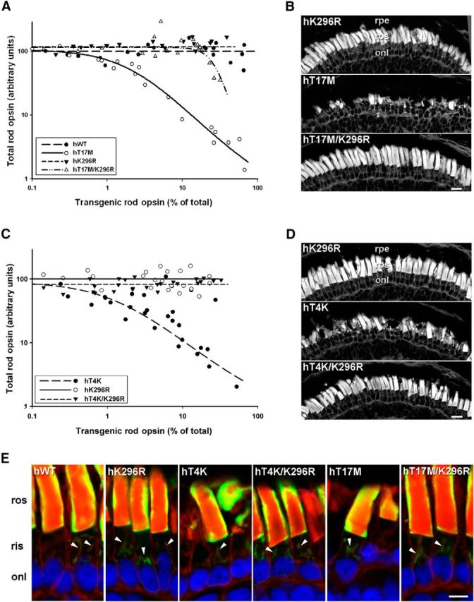
Inactive T4K and T17M opsins exhibited reduced rod toxicity. A, C, Plots of transgenic opsin expression levels versus total rod opsin levels from primary transgenic tadpoles expressing (A) hWT, hT17M, hK296R, or hT17M/K296R rhodopsins (n = 22 per group) or (C) hT4K, hK296R, or hT4K/K296R rhodopsins (n = 29 per group). B, D, Confocal micrographs of cryosections from transgenic retinas stained with WGA. Retinas expressing hK296R, hT17M/K296R, or hT4K/K296R opsin appeared healthy with numerous long closely packed ROS. Retinas expressing hT17M or hT4K rhodopsin exhibited loss or shortening of rod OS. E, Confocal micrographs of retinal cryosections labeled with mAb 2B2 (green) and counterstained with WGA (red) and Hoechst nuclear stain (blue). All mutants (T4K, T17M and K296R, T4K/K296R, and T17M/K296R) localized to the rod OS and Golgi (arrowheads) in a pattern indistinguishable from WT. rpe, Retinal pigment epithelium; ros, rod outer segment; onl, outer nuclear layer. Scale bars: B, D, 20 μm; E, 10 μm.