
Figure 4.
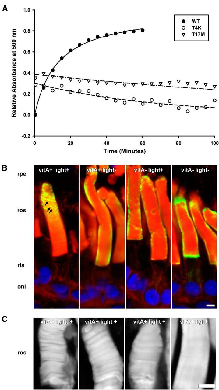
T4K and T17M rhodopsins exhibit instability in vitro and in vivo. A, Regeneration of heterologously expressed hWT, hT4K, and hT17M pigment. A500 measurements were obtained after >495 nm bleach of purified reconstituted pigments. B, C, Confocal micrographs of cryosections from transgenic retinas expressing hT4K opsin labeled with mAb 2B2 (green) and counterstained with WGA (red) and Hoechst nuclear stain (blue; B) or stained with WGA alone (C). Under conditions promoting inactive opsin (vitA− and/or light−), hT4K opsin was uniformly detected at the periphery of the rod OS as is typical for hWT opsin. However, under conditions favoring rhodopsin activation (vitA+ and light+), hT4K opsin labeling frequently appeared punctate (B, arrows). Similarly, WGA labeling of rod OS was smooth and uniform in the absence of light but nonhomogeous indicating disruption of the disk stacks in the presence of light (C). rpe, Retinal pigment epithelium; ros, rod outer segments; onl, outer nuclear layer. Scale bar, 5 μm.