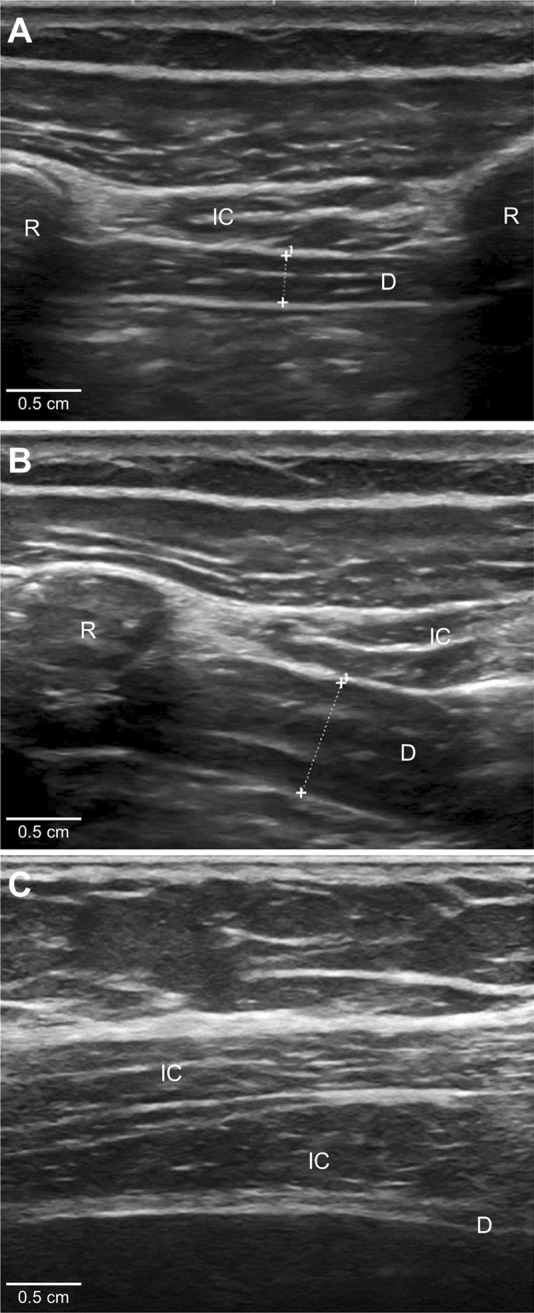
Figure 3. Ultrasound of the diaphragm.

The diaphragm (D) is the 3-layered structure situated deep to the intercostal (IC) muscles that span the 2 ribs. The diaphragm muscle tissue is hypoechoic (dark) on ultrasound, and the 2 layers of connective tissue encasing the muscle (peritoneum and parietal pleura) are hyperechoic (bright) on ultrasound. (A) A normal diaphragm at end-expiration (0.29 cm thick). (B) A normal diaphragm, in the same patient, at maximal inspiration (0.6 cm thick, giving a diaphragm thickening ratio of 2.1). (C) An atrophic diaphragm (0.05 cm thick) in a patient with phrenic neuropathy. R = rib.
