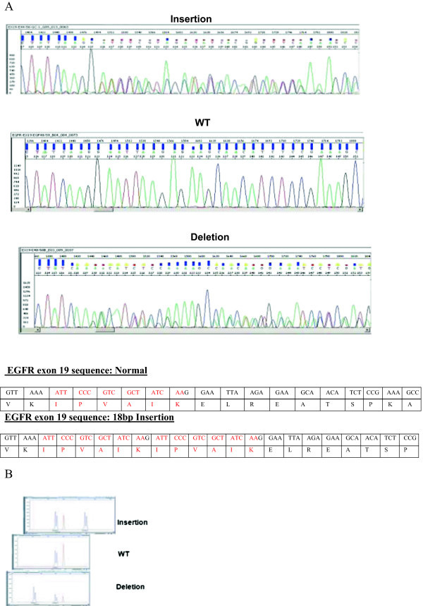
Figure 1.

Molecular analysis of the EGFR exon 19 insertion. A) Direct sequencing: Sanger sequence was performed and detected the mutation in the EGFR exon 19. Below are the electropherograms of insertion, normal (WT) and deletion sequences. The description of the normal and insertions sequences are described with their relative amino-acid. B) Fragment length analysis of EGFR exon 19: A very precise and simple visual way to detect and describe the insertion of 18 nucleotides in the EGFR exon 19. The picture shows a comparison between the insertion with the wild type and a deletion of EGFR exon 19.
