Figure 1.

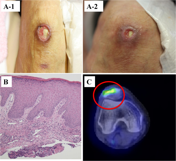
Cutaneous ulceration before and after steroid therapy; pathology and PET/CT findings of the cutaneous ulceration. (A-1) The right knee cutaneous ulcer measured 4 cm before steroid therapy. (A-2) One month after the treatment, it measured < 1 cm and showed granulation tissue growth. (B) A biopsy of the edge of the ulcer revealed a mild lymphocytic infiltrate in the superficial dermis; however, no obvious findings of vasculitis were observed. (C) FDG PET/CT demonstrated increased tracer uptake in the ulcer, suggesting inflammation.
