Fig. 3.

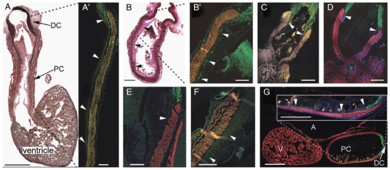
Chondrichthyans exhibit similar arterial pole architechture. (A) Sagittal section through the heart of Dalatias licha. Scale bar=5 mm. Cranial to the top, dorsal to the left—elastic trichrome stains muscle brown, collagen red, and elastic proteins black. Abundant elastic fibers in the distal smooth muscle component of the outflow tract (OFT) extend caudally into the subendocardium as far as the most proximal valve leaflets. (A′) Detail of the ventral portion of the OFT marked by the dashed lines, labeled with anti-SM22α (green). Scale bar=1 mm. Arrowheads show that smooth muscle is coincident with elastic fibers in the subendocardial layer. Autofluorescence in the myocardium has been artificially enhanced. (B) Longitudinal section through the OFT of Etmopterus spinax—elastic trichrome. Scale bar=500 μm. Subendocardial elastic fibers (arrowheads) extend to the level of the most proximal valve leaflets. (B′) Detail of the region marked by dashed lines in (B). Anti-SMA shows coincident subendocardial smooth muscle (arrowheads) in E. spinax. Scale bar=250 μm. Smooth muscle (arrowheads) extends to the most proximal valve. (C and D) The OFT of two Galeus spp., G. atlanticus, and G. melastomus, labeled with CH1 (red) and SM22α (green). Scale bars=1 mm. Smooth muscle (arrowheads) extends to the most distal valves. (E) Detail of the arterial pole of Scyliorhinus canicula. Scale bar=500 μm. The dorsal wall of the conus is shown in sagittal section. CH1 (red) and anti-SMA (green). In this species, the overlapping region is not so extensive, barely reaching the most distal valves (arrowheads). (F) Smooth muscle (arrowheads) extends beyond the distal valves in Leucoraja naevus. CH1 (red), SM22α (green). Scale bar=250 μm. (G) Sagittal section of the heart of Chimaera monstrosa—CH1 (red), SM22α (green). Scale bar=500 μm. Inset shows detail of the ventral OFT wall and that smooth muscle extends to the most proximal valves (arrowheads). Scale bar=250 μm. A, atrium; DC, distal smooth muscle component (bulbus arteriosus); PC, proximal myocardial component (conus arteriosus); V, ventricle.
