
Fig. 7.
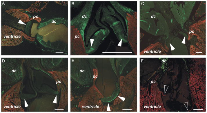
Smooth muscle and myocardium in the valve sinuses: evidence of active valving? (A–E) Five examples of teleost fish species with smooth muscle (labeled by anti-SM22α—green) in the base of the outflow valve (solid arrowheads). Myocardium is labeled by either MF20 or CH1 (red). (A) Osteoglossum bicirrhosum. (B) Channa Micropeltes. (C) Sardina pilchardus. (D) Morone chrysops. (E) Morone americana. (F) In Scomber scombrus, CH1 labeling (red) shows myocardium in the base of the valve (hollow arrowheads). DC, distal outflow tract component (bulbus arteriosus); PC, proximal outflow tract component (conus arteriosus). Scale bars=250 μm.