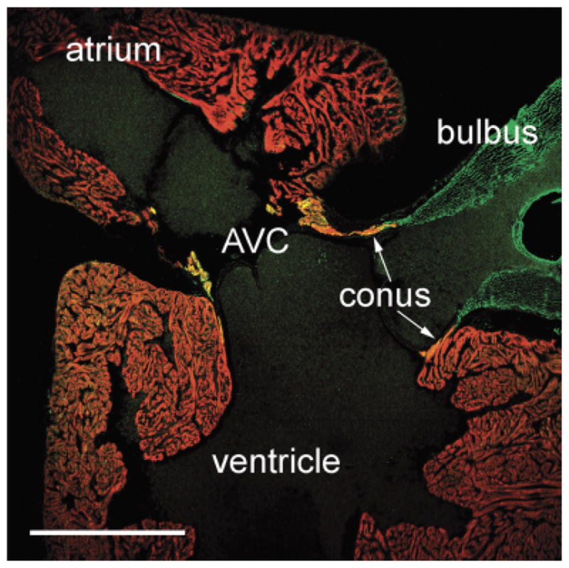
Fig. 8.

“Primitive myocardium” in the conus arteriosus and atrioventricular canal of a teleost. Sagittal section of the Atlantic silverside (Menidia menidia) heart labeled with CH1 (red) and anti-SM22α (green). Myocardium in the conus arteriosus and in the atrioventricular canal (AVC) labels positively for both antibodies (see “Discussion”). Scale bar=500 μm.
