Figure 1.
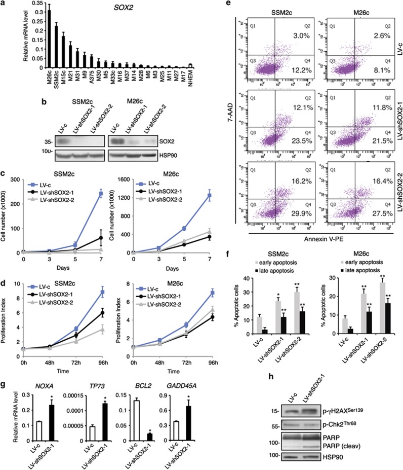
SOX2 silencing suppresses cell growth and induces apoptosis in primary melanoma cells. (a) qPCR analysis of SOX2 in a panel of 19 patient-derived melanoma cells, A375 melanoma cells and normal human epidermal melanocytes. qPCR values reflect Ct values after normalization with two housekeeping genes (GAPDH and β-ACTIN). (b) Western blotting analysis of SSM2c and M26c adherent cells stably transduced with LV-c, LV-shSOX2-1 and LV-shSOX2-2. HSP90 was used as a loading control. (c) Growth curves of SSM2c and M26c melanoma cells stably transduced with LV-c, LV-shSOX2-1 or LV-shSOX2-2. Three to five thousands transduced cells/well were plated in 12-well plates, and cells were counted on days 3, 5 and 7. (d) Proliferation index measured by CFSE staining in SSM2c and M26c melanoma cells stably transduced with LV-c, LV-shSOX2-1 or LV-shSOX2-2. (e, f) SSM2c and M26c cells transduced with LV-c, LV-shSOX2-1 or LV-shSOX2-2 were analyzed by flow cytometry for Annexin V+/7-AAD− (early apoptosis) and Annexin V+/7-AAD+ cells (late apoptosis). (g) qPCR analysis of NOXA, TP73, BCL-2 and GADD45A in M26c cells transduced with LV-c and LV-shSOX2-1. qPCR values reflect Ct values after normalization with two housekeeping genes (GAPDH and β-ACTIN). (h) Western blotting analysis of p-γH2AX, p-Chk2 and poly ADP-ribose polymerase (PARP) in M26c cells, showing increased phosphorylation of γH2AX at Ser139 and PARP cleavage in cells transfected with shSOX2 for 48 h. HSP90 was used as loading control. Data shown are the mean±s.e.m. of at least three independent experiments. *P⩽0.05; **P⩽0.01 vs LV-c.

