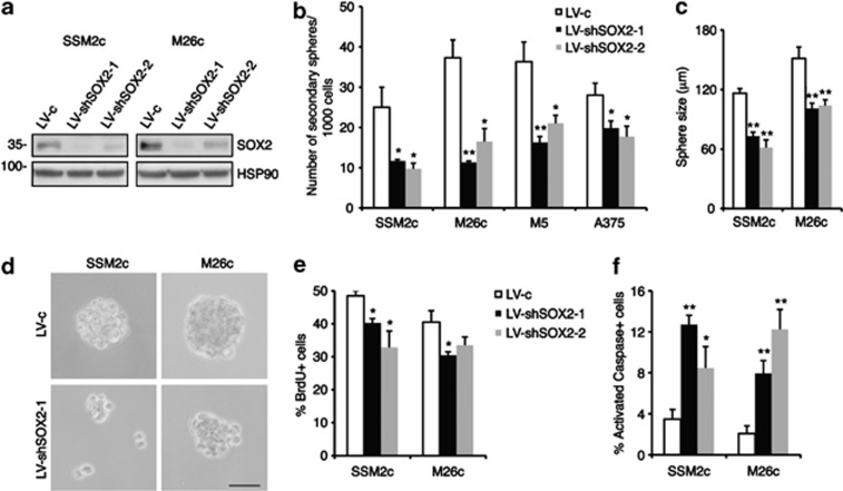
Figure 4.
Silencing of SOX2 reduces melanoma sphere self-renewal. (a) Western blotting analysis of SSM2c and M26c spheres transduced with LV-c control, LV-shSOX2-1 and LV-shSOX2-2, showing the near complete loss of SOX2 protein with both shRNAs. HSP90 was used as loading control. (b) Reduction in the number of secondary melanoma spheres after silencing of SOX2. (c) Measurement of the size of SSM2c and M26c secondary melanoma spheres after transduction with in LV-c, LV-shSOX2-1 and LV-shSOX2-2. (d) Representative images of secondary spheres as indicated in panel (c). Scale bar=150 μm. (e, f) Quantification of BrdU incorporation (e) and of activated Caspase-3+ cells (f) in SSM2c and M26c melanoma spheres after transduction with LV-c, LV-shSOX2-1 and LV-shSOX2-2. At least 10 independent fields of BrdU/DAPI and cleaved Caspase-3/DAPI-labeled cells were counted per condition. Data shown are the mean±s.e.m. of at least three independent experiments. *P⩽0.05; **P⩽0.01 vs LV-c.

