Figure 5.
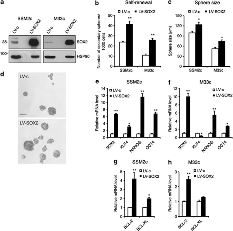
Enhanced SOX2 function increases stemness in melanoma cells. (a) Western blotting analysis shows SOX2 expression levels in control (LV-c) and SOX2-stably transfected (LV-SOX2) SSM2c and M33c melanoma cells. HSP90 was used as loading control. (b) Increase in the number of secondary spheres upon SOX2 expression. (c) Measurement of the size of secondary melanoma spheres in LV-c and LV-SOX2 transfected SSM2c and M33c spheres. (d) Representative images of secondary melanoma spheres as indicated in panel (c). Scale bar=100 μm. (e–h) Gene expression analysis of SOX2, KLF4, NANOG, OCT4 (e, f) and BCL-2 and BCL-XL (g, h) in LV-c and LV-SOX2 stably transfected melanoma spheres, as measured by qPCR. Ct values were normalized with two housekeeping genes, with the values in control spheres equated to 1. Data shown are the mean±s.e.m. of at least three independent experiments. *P⩽0.05; **P⩽0.01 vs LV-c.

