Fig. 2.

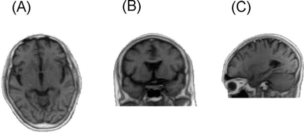
T1-weighted magnetic resonance images (MRIs) in the globus pallidus of the patient. (A) Axial T1-weighted MRI, (B) coronal T1-weighted MRI, and (C) sagittal T1-weighted MRI.

T1-weighted magnetic resonance images (MRIs) in the globus pallidus of the patient. (A) Axial T1-weighted MRI, (B) coronal T1-weighted MRI, and (C) sagittal T1-weighted MRI.