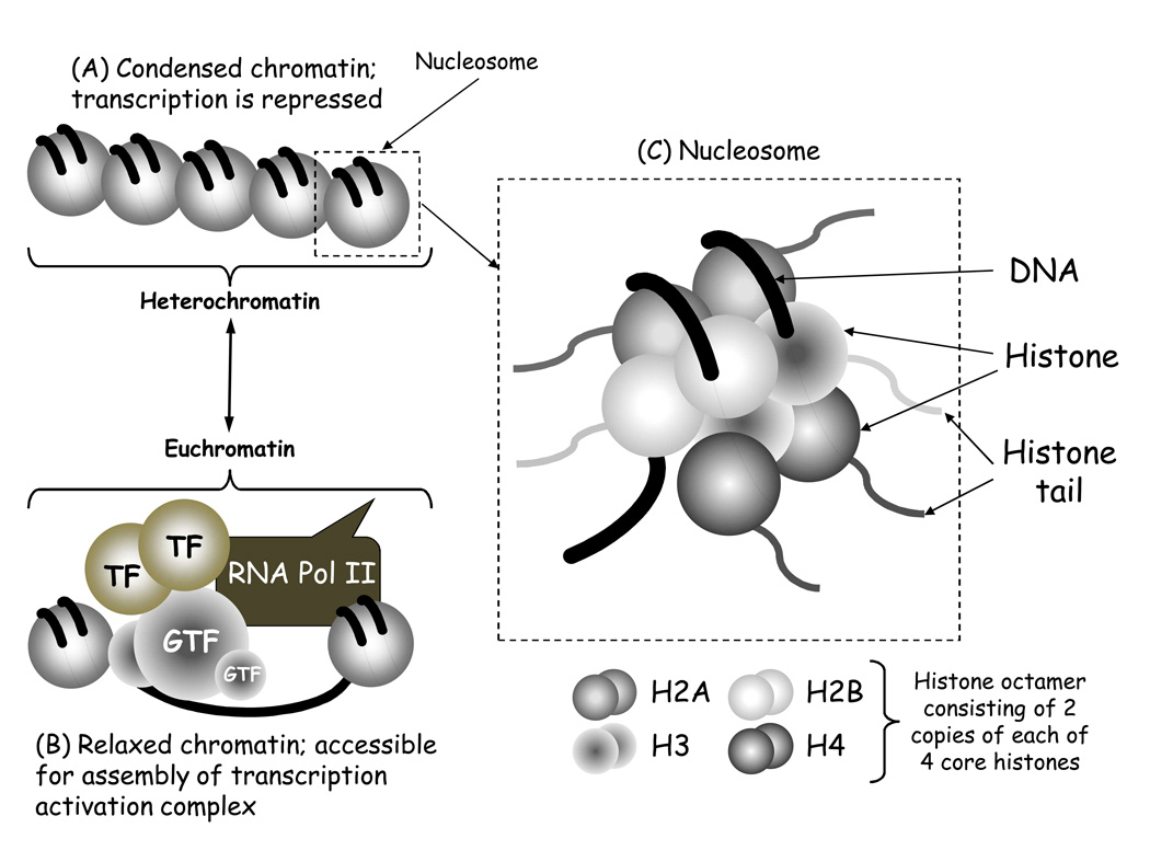
Figure 1. Diagrammatic representation of the structure of chromatin.
The classical model of chromatin structure comprises nucleosomes linked together by DNA (A). When the nucleosomes are packed tightly together chromatin is condensed and in this form transcription is repressed, and chromatin is referred to as heterochromatin. When the chromatin is modified and remodeled the nucleosomes are further apart, and in this form chromatin is relaxed making the DNA accessible to the transcription machinery (B). This allows the assembly of an active transcription complex that includes multiple general transcription factors (GTF) and inducible transcription factors (TF) as well as RNA polymerase II (RNA pol II). This form of chromatin is referred to as euchromatin. (C) Each nucleosome contains an octamer of histone proteins made up of two of each type of core histone: Histone 2A (H2A), H2B, H3 and H4. Each of these histones has an amino terminal tail that extends beyond the nucleosome structure and can be modified by chromatin enzymes.

