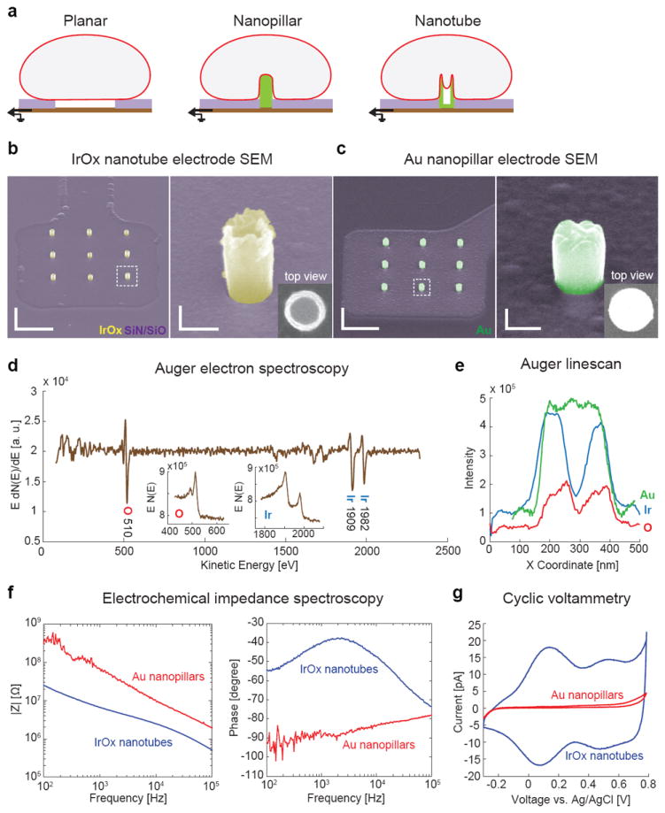
Figure 1. Characterization of vertical IrOx nanotube electrode.

a, Schematics of cells interfacing with planar, vertical nanopillar and vertical nanotube electrodes. The cell membrane not only wraps around the outside of the nanotube but also protrudes inside the hollow center. b, SEM images of a three-by-three array of IrOx nanotubes on a square Pt pad with the rest of the area insulted with Si3N4/SiO2. The hollow center of the nanotube can be clearly seen in expanded 52°-tilted view and the top view. Scale bars: left 2 μm, right 200 nm.c, SEM images of Au nanopillar electrodes show solid pillars. The diameter of the Au nanopillar is designed to be slightly larger than the IrOx nanotube so that they have similar surface area. Scale bars: left 2 μm, right 200 nm.d, Auger electron spectrum of the nanotube electrodes confirms the presence of iridium and oxygen (insets: raw spectra). e, Elemental line scans along the diameter of an IrOx nanotube and a Au nanopillar. Ir and O plots (blue and red) show two peaks at the side wall and a drop at the center, while Au (green) shows a flat top. f, Electrochemical impedance spectroscopy of IrOx nanotube and Au nanopillar electrodes in phosphate buffered saline. g, Cyclic voltammetry of IrOx nanotube and Au nanopillar electrodes in PBS at a scan rate of 30 mV/s.
