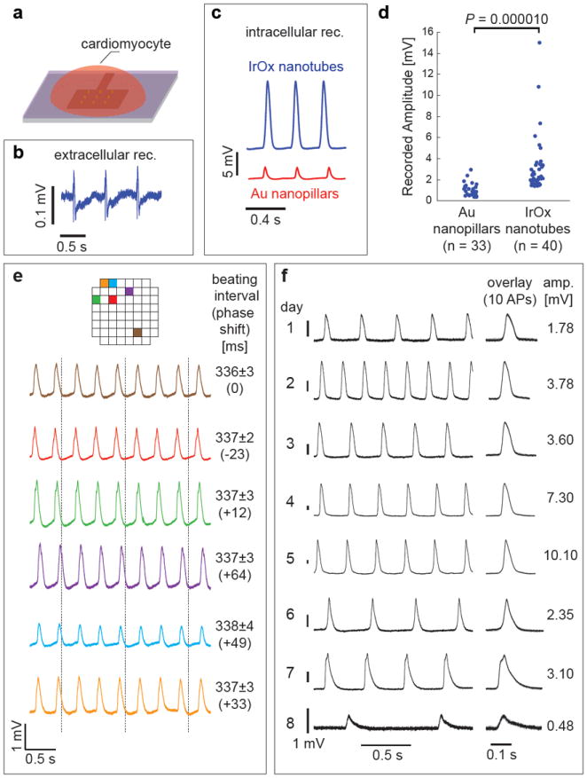
Figure 2. Intracellular recording of action potentials by the IrOx nanotube electrodes.

a, Schematic illustration of electrophysiology recording using vertical IrOx nanotube electrodes. b, Before local electroporation, nanotube electrodes measured extracellular action potentials in HL-1 cardiomyocytes. c, Immediately after local electroporation, both IrOx nanotube and Au nanopillar achieved intracellular recording of action potentials. d, Statistical measurements show that nanotube electrodes recorded much larger signal than nanopillars of similar surface area. e, Simultaneous recording by six different nanotube electrode arrays in the same culture. Adjacent array separation is 100 μm and the array positions on the chip are color labeled. The vertical dashed lines are guides to see the phase shift of action potentials among different electrodes. f, Intracellular recording of a single cell over eight consecutive days (the lifespan of the culture).
