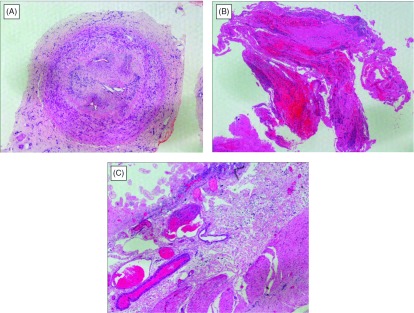
Fig. 1.

Histological findings of surgical specimens from the patient. (A) Posterior tibial artery from amputated leg contained organized thrombus. Although proliferation of fibroblasts and capillary structures with lymphocytic infiltration were totally obstructed inside of the artery, either inner or outer elastic laminae of the medial layer was intact, which distinguish thromboangiitis obliterans (TAO) for arteriosclerosis and other systemic vasculitides. Hematoxylin and eosin staining. (B) The resected specimen by embolectomy of the SMA. It comprised organized emboli mixed with fresh thrombi. Note that neutrophils infiltrates into clots. Hematoxylin and eosin staining. (C) Mucosal layer of the small intestine was extensively necrotic and villous structures were left out. Prominent congestion and edema were observed in the submucosa. There were not observed any finding of vasculitis. Hematoxylin and eosin staining, × 40.
