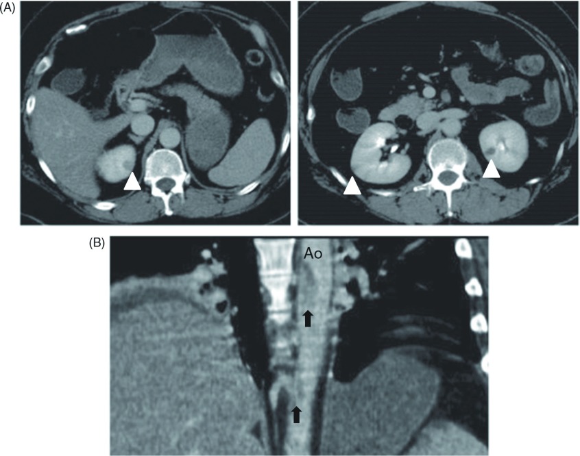
Fig. 2.
Computed tomography (CT) revealed thrombi in the descending thoracic aorta and multiple infarctions in the spleen and kidney. (A) Cross-sectional CT images showed multiple kidney infarctions (white arrowheads). (B) A sagittal CT image showed floating thrombus in the descending thoracic aorta (black arrows). Ao: aorta.

