
Figure 3. OnTEBP proteins bind ciliate telomeric ssDNA.
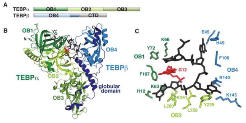
(A) Domain topology of O. nova TEBP proteins. Four OB folds are present, as well as a structurally uncharacterized C-terminal domain (CTD) in TEBPβ. (B) TEBPα OB1-3 (green) forms a complex with TEBPβ (blue) to bind ssDNA ligand (black; PDB: 2I0Q). The TEBPβ globular domain that is bound by OB3 is in dark blue. The 3′ base, G12, is colored red and is fully buried in the groove between the protein subunits. (C) OB1, OB2, and OB4 make critical contacts with the 3′ loop of the ssDNA ligand to sequester the bases from solvent.